Mole or Melanoma? Visual Signs AI Diagnostic Apps Miss Across Diverse Skin Tones

Millions of smartphone users point their phone cameras at suspicious blemishes each month hoping for immediate clarity. Instead of booking an appointment at a nearby clinic, patients frequently lean on consumer screening applications to decide whether an irregular patch warrants a doctor's visit. That shortcut can carry fatal consequences. Investigative findings published in a Civio report revealed that prominent commercial algorithms misclassify one out of every three melanomas, with diagnostic accuracy dropping precipitously whenever software assesses non-white patients.
The convenience of an on-demand smartphone scan conceals a glaring engineering blind spot. While machine learning developers trained their computer vision models on millions of digital images, those training repositories drew overwhelmingly from light-skinned populations across northern Europe and North America. For anyone typing "dermatologos cerca de mi" into a search bar seeking reliable answers about a dark streak beneath a thumbnail or a shifting patch of pigment on an ankle, trusting an algorithmic verdict introduces unacceptable clinical risk.
📌 Key Takeaways:
- Algorithmic Blind Spot: Independent evaluations reveal consumer AI vision software fails to detect approximately **33% of melanomas**, with error rates spiking dramatically on darker skin tones (Fitzpatrick types IV through VI).
- The Training Data Void: More than **80% of open-access dermatology image archives** consist of fair-skinned lesions, leaving clinical diagnostic software largely blind to subungual pigmentation and unusual lesion borders.
- Clinical Necessity: Subtle malignancies like acral lentiginous melanoma demand polarized light, direct dermoscopy examination, and tissue pathology that no mobile camera sensor can replicate.
How Training Data Created an Algorithmic Blind Spot
Machine learning models depend strictly on the historical examples fed into their neural networks. When academic labs and health-tech commercial startups gathered baseline images for computer vision pipelines, they relied on publicly accessible repositories like the International Skin Imaging Collaboration (ISIC). Independent audits across clinical informatics literature indicate that fewer than 5% of archived training photographs captured lesions on deep brown or black skin.
This demographic imbalance skews how software evaluates common signs of disease. Convolutional neural networks evaluate pigment density, peripheral borders, and contrast against surrounding skin tissue. On fair skin, an evolving melanoma frequently appears as an asymmetric, dark blemish set against a pale background. The software learns this contrast pattern reliably.
When applied to Fitzpatrick skin types V and VI, background melanin flattens the optical contrast that algorithms depend on to mark irregular boundaries. Benign seborrheic keratoses or ordinary hyperpigmentation get flagged as critical emergencies, while malignant lesions get dismissed as harmless variations. The resulting false reassurance delays clinical evaluation until tumors invade deeper vascular layers.
Why Visual Screening Rules Fail on Darker Complexions
Standard public health campaigns instruct patients to watch for the conventional ABCDE criteria: Asymmetry, Border irregularity, Color variation, Diameter greater than six millimeters, and Evolving shape. While valuable for superficial spreading melanoma on sun-exposed shoulders or forearms, this framework poorly reflects how deadly skin cancers present across diverse populations.
Patients with brown and black skin contract cutaneous malignancies far less frequently than fair-skinned individuals, but they face significantly higher mortality rates upon diagnosis. The five-year survival rate for Black patients diagnosed with melanoma stands at roughly 70%, compared to more than 90% for white patients, according to American Cancer Society registries. This survival gap stems almost entirely from delayed discovery.
In deeply pigmented skin, melanomas appear disproportionately on non-sun-exposed surfaces. Acral lentiginous melanoma (ALM), the subtype that caused the death of reggae icon Bob Marley at age thirty-six, develops on the palms of the hands, the soles of the feet, and beneath the nail beds. Mobile diagnostic tools looking for jagged sunspots routinely fail to recognize ALM in its early, treatable radial growth phase.
| Diagnostic Method | Melanoma Miss Rate | Efficacy on Types IV, VI Skin | Structural Tissue Evaluation |
|---|---|---|---|
| Consumer AI Smartphone Apps | Up to 33% (1 in 3 missed) | Very Low (uncalibrated contrast) | None (surface pixels only) |
| In-Person Clinical Dermoscopy | Approximately 5%, 10% | High (polarized cross-illumination) | Visualizes dermo-epidermal junction |
| Histopathological Tissue Biopsy | Less than 1% (Gold Standard) | Universal (independent of skin color) | Microscopic cellular architecture |
Optical Limitations: Why Phone Lenses Cannot Match a Dermatoscope
Even the most sophisticated software models face hardware limitations when processing consumer phone pictures. Standard smartphone cameras capture two-dimensional ambient light reflected off the stratum corneum, the outermost layer of dead skin cells. Surface glare, flash reflection, and post-processing software filters obscure the structural clues clinicians require to spot early malignant transformation.
A specialist uses a handheld optical instrument called a dermatoscope. This device combines high-magnification lenses with cross-polarized illumination, eliminating surface glare and rendering the upper layers of skin translucent.
Through polarized dermoscopy, a physician looks beneath the skin surface to evaluate:
- Whether melanin networks show delicate reticular regularity or disjointed, thickened lines.
- The precise architecture of microcapillaries feeding the lesion, including hairpin, dotted, or irregular vessels.
- Sub-Surface Deposits: Crystalline structures, chrysalis streaks, and deep blue-white veils indicative of invasive dermal growth.
- The parallel furrow pattern typical of benign acral moles versus the parallel ridge pattern indicative of early acral melanoma.
No consumer phone camera captures these subsurface optical features. A smartphone screen simply records a brown or black blob, leaving software algorithms to guess what lies beneath.
The Danger of Replacing Physical Checkups with App Scans
When patients notice an atypical spot, digital friction often shapes their choices. Navigating insurance directories, booking appointments, and waiting weeks for an open slot creates frustration. By contrast, a mobile scanner promises an answer in five seconds.
That dynamic creates a false sense of security. A patient who scans an atypical foot lesion and receives a "low-risk" digital score often skips physical evaluation entirely. When the spot later ulcerates or spreads into regional lymph nodes, the therapeutic window for simple surgical excision has already closed.
Bilingual and immigrant households seeking nearby care routinely query local terms like "dermatologos cerca de mi" or "melanoma screening near me" to locate trusted regional clinicians. Navigating language barriers and appointment scheduling presents real administrative friction, but skipping that search in favor of an unvalidated mobile application carries profound risks. The price of an erroneous app notification is measured in delayed treatment, radical excisions, and systemic chemotherapy.
Navigating Your Clinical Visit: What to Expect During an Exam
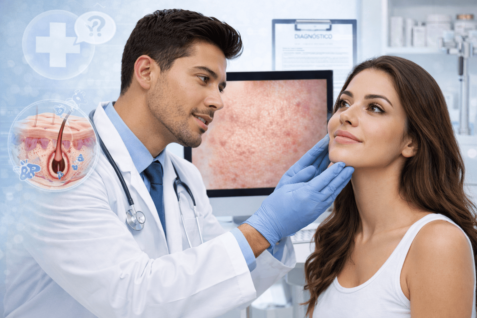
Scheduling an evaluation at an accredited medical practice replaces algorithmic guesswork with precise anatomical assessment. During an in-person full-body check, a practitioner inspects the entire cutaneous surface from scalp to toes, including skin folds, webbing between fingers, and the soles of the feet.
If a practitioner spots a suspicious mole evaluation target, the visit proceeds through structured diagnostic steps:
- High-Magnification Dermoscopic Assessment: The physician examines structural symmetry, reticular lines, and blood flow paths through specialized polarized optics.
- Clinical History Integration: The specialist accounts for personal risk metrics, including family history, past dysplastic nevi, immunosuppression, and how long the spot has been changing.
- Skin Biopsy Procedure: When a lesion exhibits suspect architecture, the area is numbed with local anesthetic. A precise punch, shave, or excisional biopsy removes tissue for laboratory evaluation.
- Pathological Staging: A dermatopathologist slices, stains, and microscopically reviews the cells to establish an exact diagnosis, measuring thickness down to hundredths of a millimeter (Breslow depth).
Direct tissue pathology remains the definitive medical benchmark. No machine learning application can extract cellular architecture from a smartphone snapshot.
Frequently Asked Questions (FAQ)
Q1: How does a suspicious mole on darker skin typically look compared to lighter skin?
A1: While fair-skinned individuals frequently develop superficial spreading melanomas on sun-exposed areas like the back and arms, patients with deeper complexions more often develop lesions on non-sun-exposed regions. These include the palms, soles of the feet, mucosal membranes, and beneath fingernails or toenails (acral lentiginous melanoma). They often appear as dark vertical bands under nails or irregular brownish-black patches on the feet.
Q2: Why do AI skin screening apps struggle so significantly with Fitzpatrick types IV through VI?
A2: Computer vision systems are heavily reliant on the training datasets used to build them. Major dermatology image databases contain disproportionately high percentages of fair-skinned individuals. Furthermore, higher natural melanin content flattens the visual contrast boundaries that image analysis tools use to detect asymmetry and irregular margins.
Q3: How often should adults undergo a professional full-body skin exam?
A3: Clinical guidelines suggest an annual full-body skin check for adults with elevated risk profiles, numerous moles, or a family history of skin cancer. Individuals without elevated risk factors should still seek immediate evaluation whenever they spot a single blemish that bleeds, tingles, changes shape, or differs noticeably from all other moles on their body.
Choosing Direct Medical Evaluation Over Uncalibrated Apps
Mobile devices excel at tracking calendar alerts and logging changes in mole dimensions over time. They cannot, however, replace surgical acumen, polarized optics, and histology. The systemic diagnostic errors documented across independent health investigations underscore the hazard of relying on smartphone software to evaluate atypical lesions, especially for non-white patients.
Early-stage cutaneous melanoma carries a cure rate exceeding 98% when identified and excised before invading deeper dermal layers. Reaching that outcome requires early discovery through cross-polarized dermoscopy and skilled physical inspection. For anyone tracking a suspicious spot, the safest next step is not downloading another algorithmic scanner, but booking an evaluation with a qualified specialist at a local clinic.