Complete Guide to Rales Breath Sounds: Causes, Symptoms, and Care
A clinician presses a cold stethoscope diaphragm against a patient’s posterior chest wall, instructing them to inhale deeply through an open mouth. Beneath the surface, instead of the whisper of laminar airflow, a series of brief, explosive pops breaks through the rhythm. These sounds, historically known as rales and modernly referred to as crackles, represent one of the most critical physical findings in pulmonary medicine. As clinical reporting from the healthcentral.com Report demonstrates, separating crackling from wheezing and rhonchi is essential for pinpointing whether a patient faces acute heart failure, progressive emphysema, or an infectious consolidation.
Misinterpreting these abnormal breath sounds carries steep risks. A misdiagnosis can delay diuretic therapy in a cardiac patient or trigger inappropriate antibiotic regimens in someone experiencing pulmonary fibrosis. Understanding the acoustic mechanics of alveolar fluid, small-airway dynamics, and sound classification bridges the gap between basic bedside examination and life-saving intervention.
📌 Key Takeaways:
- Acoustic Mechanism: Fine crackles occur when micro-airways snap open during inspiration; coarse rales stem from air boluses bubbling through liquid in larger airways.
- Primary Pathologies: Left-sided congestive heart failure and pneumonia remain the primary triggers for wet crackles, whereas interstitial fibrosis yields dry, velcro-like sounds.
- Auscultation Protocol: Systematic stethoscope lung assessment requires comparing bilateral lung fields from the lung apices down to the bases during full respiratory cycles.
The Physics Behind the Crackle in Alveolar Tissue
Rales are discontinuous, non-musical adventitious breath sounds. For decades, medical dogma held that rales were purely the auditory friction of air moving through secretions. High-speed acoustic analysis paints a different picture.
Fine crackles are micro-explosions. When alveoli and tiny terminal bronchioles collapse due to elevated surface tension, interstitial fluid, or fibrotic stiffening, they seal shut during exhalation. As inspiration generates sufficient negative intrathoracic pressure, these small conduits snap open abruptly. That sudden equalization of gas pressure vibrates the surrounding parenchyma, producing a series of brief, high-frequency acoustic spikes. These sounds last roughly 5 milliseconds and mimic salt dropped onto a hot frying pan or hair rolled between two fingers close to the ear.
Coarse rales operate on a fluid-displacement principle. When inflammatory exudate, transudate, or mucus pools inside segmental bronchi and larger bronchioles, incoming air boluses physically rupture through the liquid barriers. These interruptions yield lower-pitched, louder, and longer-lasting sounds, averaging 15 milliseconds in duration. They produce a bubbling, gurgling quality often audible across both inspiration and expiration.
Acoustic Profiles: Fine Crackles, Coarse Rales, and Rhonchi
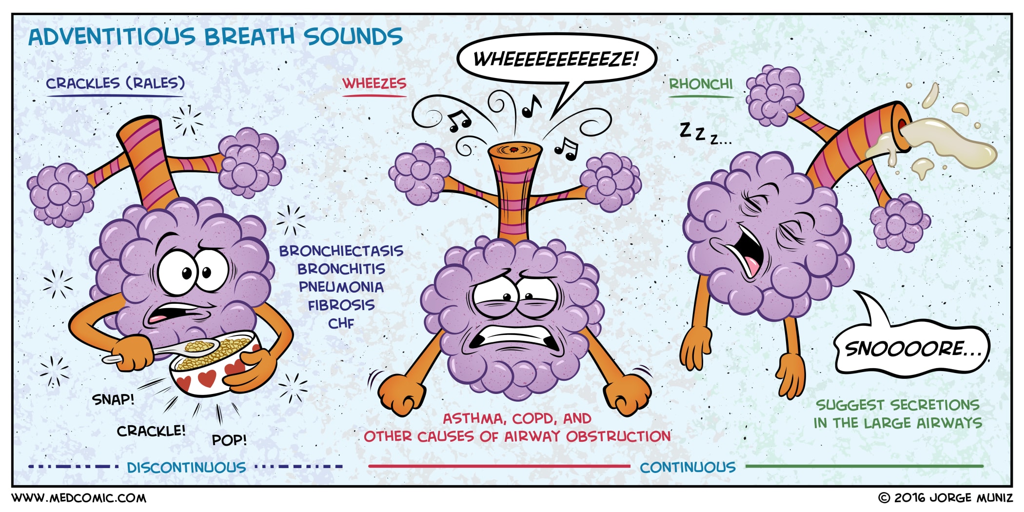
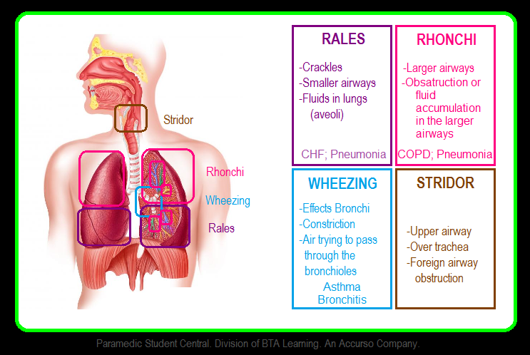
Bedside confusion between rales and other abnormal breath sounds remains common. Differentiating fine crackles from coarse variants and low-pitched rhonchi dictates whether therapy focuses on diuresis, airway clearance, or antimicrobial treatment.
| Acoustic Parameter | Fine Crackles | Coarse Rales | Rhonchi |
|---|---|---|---|
| Pitch & Timbre | High-pitched; dry, crisp, popping | Low-pitched; moist, bubbling | Very low-pitched; continuous, snoring |
| Respiratory Timing | Mid-to-late inspiration | Early inspiration and expiration | Predominantly expiration |
| Pathophysiology | Sudden airway reopening; micro-atelectasis | Air passing through airway fluid | Turbulent flow past airway secretions |
| Effect of Cough | Unchanged | Occasionally shifts or diminishes | Frequently clears or alters in pitch |
| Primary Examples | Pulmonary fibrosis, early heart failure | Severe pulmonary edema, pneumonia | Acute bronchitis, chronic bronchitis |
Rhonchi vs rales is an essential distinction. Rhonchi indicate secretions obstructing larger conduct pipes. When a patient coughs vigorously, rhonchi often shift position or vanish entirely as the sputum plug dislodges. Rales arising from parenchymal remodeling or pulmonary capillary transudation remain audible through forceful coughing.
Underlying Pathologies: From Heart Failure to Pneumonia
Detecting crackles requires placing the sound within a broader diagnostic framework. The underlying trigger dictates the acuity of patient care.
In congestive heart failure, elevated hydrostatic pressure within the pulmonary capillaries forces liquid across the endothelial barrier into interstitial spaces and alveolar units. The result is cardiogenic pulmonary edema. Auscultation reveals dependent, bilateral, late-inspiratory fine-to-coarse crackles starting at the lung bases. If left ventricles continue to struggle, the fluid rises higher, producing coarse rales throughout the mid- and upper lung zones.
A pneumonia diagnosis produces an entirely different physical profile. Infectious exudate and cellular debris concentrate in localized lobes. Auscultation over the affected segment reveals unilateral, coarse rales accompanied by bronchial breath sounds and increased tactile fremitus. The sound does not shift with position changes, confirming dense tissue consolidation rather than mobile systemic fluid.
In cases of COPD exacerbation, chronic airway inflammation overlaps with acute hypersecretion. Patients present with mixed acoustic signals: expiratory wheezing alongside coarse rales and rhonchi. When severe airway narrowing occurs, vesicular sounds diminish significantly, making crackles fainter despite profound respiratory distress.
Atelectasis presents a benign, transient variation. Patients emerging from general anesthesia or prolonged supine bed rest often exhibit basilar fine crackles. Shallow breathing leaves the lung bases under-ventilated, causing partial collapse of dependent alveoli. These crackles typically clear completely after five to ten deep breaths or sustained incentive spirometry maneuvers.
Stethoscope Lung Assessment and Modern Acoustic Triage
Precise lung auscultation requires deliberate technique. Clinicians must warm the stethoscope diaphragm and place it firmly against bare skin; auscultating through hospital gowns or clothing introduces friction artifacts that mimic rales.
The patient sits upright while taking slow, deep inspirations through an open mouth. Auscultation begins at the lung apices above the clavicles, progressing downward across the intercostal spaces in a ladder-pattern comparison: right side, then matching left side. Clinicians evaluate at least six symmetric posterior zones, four anterior zones, and two lateral mid-axillary points.
Documenting the sound requires specifying:
- Phase: Early, mid, or late inspiratory vs. expiratory.
- Pitch: High-frequency fine versus low-frequency coarse.
- Distribution: Unilateral lobar focus versus bilateral base presentation.
- Persistence: Cleared by coughing or remaining static across serial exams.
Bedside evaluation is also advancing beyond manual stethoscopes. Continuous acoustic monitoring is entering modern critical care. According to clinical trial findings reported by Medical Xpress, non-invasive digital biosensors can continuously track adventitious breath sounds to accurately predict post-extubation respiratory failure in intensive care units. These automated systems detect micro-acoustic changes hours before arterial blood oxygen levels drop, alerting medical teams to fluid shifts or airway collapse before visual distress appears.
Targeted Clinical Management and Therapeutic Protocols
Rales are acoustic warnings, not diagnoses. Treatment must target the underlying driver rather than suppressing the noise itself.
Cardiogenic pulmonary edema demands rapid volume reduction and preload reduction. Intravenous loop diuretics like furosemide drop hydrostatic pressure, while supplementary oxygen or continuous positive airway pressure (CPAP) keeps fluid-laden alveoli recruited. Vasodilators such as nitroglycerin reduce cardiac afterload in hypertensive crises, quickly reducing pulmonary congestion.
Bacterial pneumonia requires targeted antimicrobial therapy based on community-acquired or hospital-acquired risk stratification. Supplemental humidified oxygen helps alleviate hypoxemia, while chest physiotherapy aids in mobilizing thick endobronchial exudates.
For COPD exacerbations featuring rales, therapy shifts to inhaled short-acting beta-2 agonists combined with anticholinergics to open constricted passages, oral or intravenous systemic corticosteroids to reduce mucosal swelling, and controlled supplemental oxygen. Clinicians avoid high-flow unmonitored oxygen to prevent hypercapnic respiratory failure.
When crackles reflect passive atelectasis, pharmacological intervention is unnecessary. Early ambulation, routine deep-breathing exercises, and flutter-valve devices re-expand collapsed lung tissue and eliminate abnormal sounds within hours.
Frequently Asked Questions (FAQ)
Can rales breath sounds be heard without a stethoscope?
Severe coarse rales, sometimes described as a "death rattle" in end-stage secretions or intense pulmonary edema, can occasionally be heard by the unaided ear at the bedside. Most fine and coarse crackles produce micro-acoustic waves that require a stethoscope placed against the thoracic wall for detection.
How can clinicians quickly tell rales apart from pleural friction rubs?
A pleural friction rub produces a harsh, creaking, or grating sound caused by inflamed visceral and parietal pleura sliding against each other. Unlike rales, a pleural rub sounds exceptionally superficial, occurs during both inspiration and expiration with equal intensity, and vanishes if the patient temporarily holds their breath.
Are rales always a sign of a critical medical condition?
No. Transient fine crackles often appear in healthy older adults or bedbound individuals after periods of shallow breathing or prolonged sleep. These physiological atelectatic crackles clear rapidly after a few deep, forced breaths or deliberate coughs.
Refining Respiratory Diagnostics in Clinical Practice
Recognizing rales breath sounds remains a core clinical skill. While chest radiography, point-of-care lung ultrasound, and arterial blood gas analysis provide crucial diagnostic confirmation, physical auscultation remains the fastest bedside assessment available. Correctly distinguishing fine crackles from coarse rales and sonorous rhonchi guides clinicians directly toward the underlying problem, whether that means adjusting diuresis for heart failure, starting targeted antibiotics for pneumonia, or mobilizing patients to resolve atelectasis. Mastering these subtle acoustic cues ensures prompt, accurate treatment when every breath counts.