Redness vs. Induration: The Critical Mistake People Make Reading a TB Skin Test
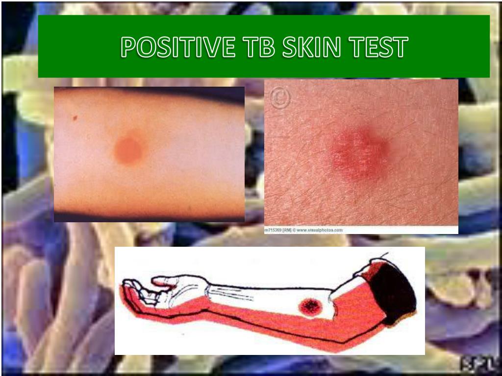
Every year, millions of healthcare workers, teachers, and international travelers roll up their sleeves for a Mantoux tuberculin skin test, only to panic two days later over a patch of red skin. Clinical field reports and ongoing surveillance from the Infectious Disease Special Edition Report reveal how frequently cutaneous hypersensitivity reactions trigger false alarms during routine screenings. The widespread instinct is to glance down at an angry red circle and assume infection, but that visual check is fundamentally flawed.
A positive tuberculosis skin test is determined entirely by touch, not color. What clinicians search for during the standardized 48 to 72 hour reading window is induration: a firm, dense, raised swelling beneath the epidermal layer. Flat surface discoloration, known as erythema, is clinically irrelevant. Misinterpreting normal irritation as active infection sends thousands into unnecessary spirals of anxiety, while missing faint, deep swelling risks leaving a dangerous pathogen undetected.
📌 Key Takeaways:
- The Core Diagnostic Rule: A positive result requires palpable induration, a dense, hard raised bump measurement, rather than flat surface redness (erythema).
- The 48 to 72 Hour Window: Reading the forearm test before 48 hours or after 72 hours invalidates the result, as delayed-type hypersensitivity peaks strictly during this timeframe.
- Variable Cutoffs: The millimeter threshold cutoff changes based on individual immune status, spanning 5 mm, 10 mm, or 15 mm depending on specific risk profiles.
- Next Diagnostic Steps: A reactive skin test does not confirm contagious illness; clinicians require a follow-up chest X-ray confirmation and an IGRA blood draw to distinguish latent infection from active disease.
Erythema vs. Induration: The Mistake That Drives False Alarms
The most common point of confusion among patients monitoring their forearm after a purified protein derivative (PPD) injection is the visible red halo. The forearm may look irritated, inflamed, or violently pink, yet remain completely negative. Erythema is simply a localized histamine reaction to the mechanical needle puncture or the carrier fluid. It carries zero diagnostic weight in tuberculosis screening.
Diagnostic confirmation rests exclusively on PPD skin test induration. Induration feels like a hard, rubbery disc embedded beneath the surface of the skin, akin to a small nickel or pencil eraser wedged under the forearm tissue. When trained nurses evaluate the site, they close their eyes or look away, sweeping fingertips lightly across the injection plane to feel where the skin changes density.
A flat, crimson splotch measuring three centimeters across is clinically negative if the skin remains pliable and soft. Conversely, a faint, flesh-colored elevation measuring 12 millimeters that feels firm under direct palpation represents a positive Mantoux test. Gauging the result visually with a ruler without feeling the margin produces misdiagnoses every single week.
The Millimeter Threshold Cutoffs: Why One Size Does Not Fit All
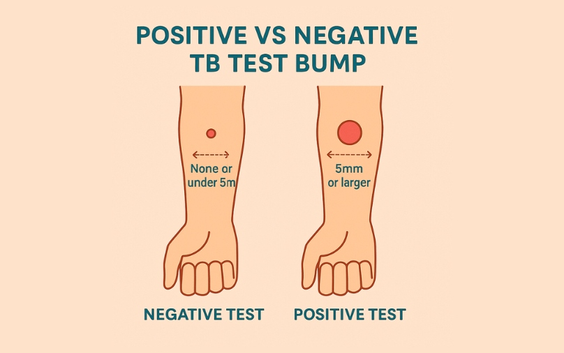
A universal number for a positive result does not exist. The Centers for Disease Control and Prevention (CDC) enforce a stratified scale that adjusts the diagnostic threshold according to the patient's immune competence and exposure probability. What marks an alarming reaction in an immunocompromised individual might register as entirely negative in a healthy adult with no known exposures.
Measurement focuses exclusively on the transverse diameter, the distance across the arm perpendicular to the long axis of the forearm. Longitudinal measurements along the length of the arm are ignored.
| Cutoff Threshold | Qualifying Patient Demographics | Underlying Clinical Rationale |
|---|---|---|
| 5 mm or greater | People living with HIV, recent contacts of infectious TB patients, organ transplant recipients, and individuals taking ≥15 mg/day of prednisone or TNF-alpha antagonists. | Severe immunosuppressed risk factors blunt the T-cell response, meaning even a small physical bump signals true exposure. |
| 10 mm or greater | Recent arrivals (within 5 years) from high-prevalence countries, injection drug users, mycobacteriology laboratory personnel, healthcare workers, and high-risk congregate setting residents. | Moderate occupational or environmental exposure risk warrants a balanced threshold to detect genuine transmission. |
| 15 mm or greater | Individuals with no known risk factors for tuberculosis, including routine workplace entrants without high-exposure duties. | Strict cutoff prevents unnecessary therapy driven by non-specific cross-reactivity in zero-risk cohorts. |
For an organ transplant recipient on heavy immunosuppressants, a firm bump measuring barely 6 millimeters signals infection. If an office worker with no clinical risk factors displays that exact same measurement, the test is documented as negative.
The Strict Mechanics of the 48 to 72 Hour Reading Window
The biochemical process behind the Mantoux tuberculin skin test is a Type IV cell-mediated delayed hypersensitivity reaction. When the diagnostic antigen is injected intradermally, sensitized memory T lymphocytes do not arrive immediately. They must recognize the antigen, migrate through capillary beds to the forearm, and release inflammatory cytokines that recruit mononuclear cells.
This cellular accumulation reaches its physical peak between 48 and 72 hours after administration. If a patient examines the site at 24 hours, normal localized trauma might present as an intimidating, tender swelling that fades completely by the 48-hour mark. Reading the test early yields frequent false positives based on simple injection trauma.
Checking the forearm past 72 hours introduces the opposite hazard. After three full days, the cellular infiltration naturally dissipates. A true 12-millimeter induration can soften, shrink below the threshold, and lead to an unrecorded positive. If an appointment is missed and the window lapses, clinical protocols dictate that the test must be repeated from scratch on the opposite arm.
False Positives, the BCG Vaccine, and the Role of Blood Tests
One of the largest sources of confusion in modern screening involves childhood vaccinations. Many individuals born outside the United States received the Bacille Calmette-Guérin (BCG) vaccine during infancy to protect against severe pediatric tuberculous meningitis. The BCG formulation relies on an attenuated strain of Mycobacterium bovis, which shares multiple cross-reactive antigens with Mycobacterium tuberculosis.
As a result, a healthy person who received the BCG vaccine years prior will frequently display a hard, raised induration measuring 10 to 14 millimeters during a Mantoux test, creating a false-positive result. Their immune system is responding to the ancestral vaccine antigens, not a wild-type infection.
Diagnostic Pathway Following Induration:
[Palpable Induration Detected]
│
├── Check BCG History / Potential Non-TB Mycobacteria
│ │
│ └── Run TB Blood Test (IGRA: QuantiFERON / T-SPOT)
│ │
│ ├── IGRA Negative: False positive skin reaction ruled out
│ └── IGRA Positive: Infection confirmed
│
└── Mandatory Chest X-Ray Confirmation
│
├── Clear Lungs + Asymptomatic: Latent TB Diagnosis
└── Cavitary Lesions / Infiltrates: Active Pulmonary Tuberculosis
To resolve this ambiguity, modern diagnostic protocols employ an interferon-gamma release assay (TB blood test IGRA), marketed under names like QuantiFERON-TB Gold Plus and T-SPOT.TB. These laboratory assays measure how laboratory-mixed T-cells respond to distinct genetic proteins, specifically ESAT-6 and CFP-10, that exist inside Mycobacterium tuberculosis but are absent from the BCG vaccine strain and most environmental non-tuberculous mycobacteria. When skin tests yield ambiguous or BCG-clouded bumps, the blood test serves as the ultimate arbiter.
Latent Infection vs. Active Pulmonary Tuberculosis: Understanding the Difference
Discovering a confirmed positive bump does not mean someone is coughing up bacteria or endangering family members. A positive Mantoux test merely establishes that the immune system has encountered the tuberculosis organism at some point in the patient's life. It does not measure infectiousness.
The critical separation lies between latent tuberculosis diagnosis and active pulmonary tuberculosis:
- Latent TB Infection: The immune system has successfully walled off the bacteria inside microscopic granulomas. Patients carry zero symptoms, feel completely healthy, cannot transmit the bacteria to anyone else, and show completely clear lung imaging. However, the bacteria remain dormant and retain roughly a 5% to 10% lifetime risk of reactivating if immune defenses slip.
- Active TB Disease: The immune barrier collapses, allowing the bacilli to multiply aggressively. Patients suffer systemic symptoms like night sweats, unexplained weight loss, persistent productive cough lasting over three weeks, and hemoptysis (coughing up blood). In this state, the patient is highly infectious and requires urgent, multi-agent antibiotic therapy.
A chest X-ray confirmation represents the non-negotiable step following any positive skin test. Radiologists scan the pulmonary fields for apical infiltrates, cavitary lesions, or calcified Ghon complexes. If the chest radiograph returns completely normal and the patient shows no systemic symptoms, the diagnosis is confirmed as latent tuberculosis, treated preventatively with regimens such as weekly isoniazid and rifapentine for three months.
When to Seek Urgent Clinical Evaluation
While a routine positive skin test requires calm scheduling for an X-ray and physician consult, certain physical reactions warrant fast intervention. Severe hypersensitivity can occasionally trigger blistering, skin sloughing, or ulceration directly over the forearm injection site. These reactions indicate an intense cellular response and must be evaluated by a healthcare practitioner right away.
Immediate clinical escalation is required if any of the following signs appear alongside a developing skin reaction:
- Unexplained coughing that has persisted longer than three weeks, particularly with blood in the sputum.
- High fever, severe chills, and drenching sweats that soak through clothing overnight.
- Sudden shortness of breath or localized pleuritic chest pain.
- Severe cutaneous necrosis or open ulceration spreading across the forearm injection site.
For everyday screenings, remember the primary physical rule: close your eyes, lightly slide your fingers across the forearm, and ignore the redness. If there is no firm, raised disc beneath the surface at hour 48, that wide red mark is likely nothing more than an irritated skin patch.
Frequently Asked Questions (FAQ)
Q1: What does a negative TB skin test look like?
A1: A negative test typically shows flat, smooth skin at the injection site. There may be a faint red spot from the needle poke or a broad patch of light pink redness, but running your fingers across the skin reveals no dense, hard bump beneath the surface.
Q2: Can I scratch my arm if the test site starts itching?
A2: No. Scratching can trigger localized swelling, introduce bacteria, and cause mechanical inflammation that makes accurate clinical measurement difficult. If the site itches, apply a cool, damp washcloth without rubbing.
Q3: Will a positive TB skin test ever go back to being negative later in life?
A3: Generally, no. Once your immune system produces memory T-cells sensitized to tuberculin antigens, you will almost always test positive on future skin tests. For this reason, individuals with a documented positive should not receive annual Mantoux tests; employers rely on symptom questionnaires and baseline chest X-rays instead.
Q4: Does bruising at the injection site mean the test is positive?
A4: Bruising does not indicate a positive result. Bruising happens when the small intradermal needle nicks a microscopic capillary during injection. A bruise is flat tissue discoloration and carries no diagnostic significance without an underlying firm induration.
Navigating Your Results with Confidence
Facing a suspicious reaction on your forearm can be unsettling, but clinical context is paramount. A positive result on a Mantoux tuberculin skin test is not a sentence of acute illness; it is an early-warning signal that your immune system carries immunological memory of an exposure. Modern medicine possesses exceptionally precise tools, from IGRA blood assays to targeted short-course preventative antibiotics, designed to neutralize latent organisms long before they develop into active disease.
Step away from magnifying mirrors and harsh bathroom lighting. Allow trained clinical staff to palpate the site within the verified 48 to 72 hour reading window. Relying on professional measurement of palpable induration rather than surface color ensures you avoid needless panic while taking appropriate, measured steps toward your long-term health.