Stroke vs Aneurysm on Scans: How Doctors Spot the Difference Under Pressure
When a patient arrives at an emergency department with acute neurological collapse, minutes dictate whether brain tissue survives or dies. The clinical symptoms can look deceptively similar: slurred speech, sudden weakness, disorientation, or an incapacitating headache. Yet the underlying pathology often belongs to two distinct vascular crises: an ischemic stroke or a ruptured brain aneurysm producing a subarachnoid hemorrhage. Confusing the two is catastrophic. Administering clot-busting medication to an actively bleeding aneurysm can prove fatal, while delaying revascularization during an ischemic blockage destroys millions of neurons every minute.
Recent clinical presentations at major stroke assemblies underscore how rapidly the imaging landscape is shifting. As documented in a NeuroNews International Report covering late-breaking data from the International Stroke Conference, automated detection models and advanced emergency radiology triage software are streamlining the critical window between hospital arrival and targeted intervention. Understanding the radiological markers that differentiate these conditions reveals how neurovascular teams read subtle variations in tissue density, vessel geometry, and fluid movement to make split-second therapeutic choices.
📌 Key Takeaways:
- The Immediate Distinction: An ischemic stroke involves an obstructed artery choking cerebral blood flow, while an unruptured intracranial aneurysm is a structural outpouching that becomes a hemorrhagic emergency only upon rupture.
- The Diagnostic Protocol: Emergency teams run a non-contrast head CT first to rule out active hemorrhage before considering thrombolysis, moving immediately to CT angiography to pinpoint arterial occlusions or vascular malformations.
- The Treatment Divergence: While acute ischemic blockages require clot dissolution or mechanical extraction, a ruptured aneurysm demands immediate blood pressure reduction and endovascular coiling or surgical clipping to arrest intracranial bleeding.
The First Fifteen Minutes Inside Emergency Radiology Triage
The diagnostic clock begins the second the patient enters the trauma bay. Hospital triage algorithms prioritize brain imaging over almost all other laboratory diagnostics because physical examinations alone cannot definitively reveal what is happening inside the skull. A focal motor deficit, such as unilateral arm weakness or facial droop, classically suggests an ischemic stroke, yet an intracranial aneurysm that ruptures into brain parenchyma can trigger the exact same deficits. Conversely, an ischemic occlusion at the tip of the basilar artery can produce sudden loss of consciousness that mimics a catastrophic aneurysmal bleed.
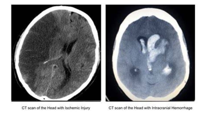
The initial baseline tool remains the non-contrast head computed tomography (NCCT) scan. It is fast, taking less than forty-five seconds of actual scan time, and universally accessible in modern stroke centers. The non-contrast CT does not immediately reveal every ischemic stroke; its paramount task is identifying extravasated blood. Blood shows up bright white on an NCCT due to the high density of hemoglobin protein globin molecules. If the scanner reveals no hyperdense pooling, the clinical pathway veers toward ischemic stroke protocols, unlocking consideration for intravenous tPA administration or tenecteplase. If high-density fluid pools within the subarachnoid spaces or deep parenchymal fissures, the pathway shifts instantly to neurosurgical aneurysm management.
Reading the Densities: How Blood and Blockages Surface on Scans
On a plain non-contrast CT, the difference between these pathologies comes down to Hounsfield units, the quantitative scale used by radiologists to measure radiodensity. Acute blood typically measures between 50 and 70 Hounsfield units, appearing as an unmistakable bright, hyperdense fluid. In a ruptured brain aneurysm, this blood escapes under arterial pressure, filling the basal cisterns, Sylvian fissures, and cortical sulci in a characteristic starburst or branched pattern known as a subarachnoid hemorrhage.
Ischemia looks entirely different. When an embolus lodges in a cerebral vessel, it abruptly halts downstream cerebral blood flow. Deprived of oxygen and glucose, cellular sodium-potassium pumps fail, causing cytotoxic edema as water rushes into swollen neurons. Water possesses a low density (zero Hounsfield units), which causes ischemic brain tissue to appear dark and hypoattenuating. However, this parenchymal hypodensity takes hours to fully manifest on a standard CT. In the earliest phase of an ischemic stroke, radiologists must hunt for subtle indirect indicators:
The "dense middle cerebral artery sign" appears when a fresh, red blood cell-rich clot sits directly within the horizontal segment of the arterial vessel, appearing hyperdense against normal circulating blood. Radiologists also look for the loss of the insular ribbon, where the crisp boundary between gray matter and white matter at the lateral cortex blurs into a uniform gray smudge due to cellular swelling. If the scan shows bright branching fluid spanning the basal cisterns rather than regional gray-white blurring, the diagnosis is a ruptured intracranial aneurysm, not an arterial occlusion.
Diagnostic Profiles and Structural Differences
Differentiating these emergencies relies on strict radiological patterns, structural vascular changes, and the exact clinical timeline documented upon arrival.
| Diagnostic Variable | Ischemic Stroke | Ruptured Intracranial Aneurysm |
|---|---|---|
| Primary Mechanism | Thromboembolic arterial occlusion | Structural dome rupture under arterial pressure |
| Hallmark Clinical Sign | Sudden painless focal neurological deficit | Instantaneous, peak-intensity thunderclap headache |
| Non-Contrast CT Findings | Early hypoattenuation or normal scan (first 3 hours) | Hyperdense (bright white) blood in basal cisterns |
| Angiography Presentation | Abrupt vessel cutoff with downstream perfusion deficit | Saccular or fusiform vascular outpouching |
| Primary Interventions | Intravenous thrombolysis or mechanical thrombectomy | Endovascular coiling, flow diversion, or surgical clipping |
| Acute Target Window | 0, 4.5 hours (tPA); up to 24 hours (thrombectomy) | Immediate exclusion to prevent fatal rebleeding within 24 hours |
CT Angiography and the Hunt for Vascular Outpouchings
Once the plain scan establishes whether blood is present, clinicians immediately transition the patient to CT angiography (CTA). By injecting an iodinated contrast agent through a peripheral intravenous line, clinicians turn the cerebral vascular tree visible under dynamic X-ray scanning. CTA bridges the gap between seeing the effect of vascular damage and identifying its precise anatomical cause.
In cases where the plain CT shows subarachnoid hemorrhage, CTA serves as the definitive anatomical roadmap for neurosurgeons. Most intracranial aneurysms arise at arterial branch points along the circle of Willis, a vascular ring at the base of the brain where hemodynamic stress is highest. The anterior communicating artery, posterior communicating artery, and the bifurcation of the middle cerebral artery account for the vast majority.
CTA reconstruction displays these structures in high-resolution three dimensions, exposing the precise morphology of the aneurysm: its neck width, dome diameter, aspect ratio, and whether secondary blebs (daughter sacs) are protruding from its walls. Identifying an irregular, multilobular dome confirms the exact site of rupture among patients who possess multiple incidental unruptured lesions.
If the non-contrast scan showed no bleeding, CTA plays the opposite role: hunting for large vessel occlusions (LVOs). Instead of looking for an abnormal bubble of blood protruding outward, the radiologist tracks the flow of contrast down major channels like the internal carotid or middle cerebral arteries, looking for an abrupt, square-edged stop where an embolus has blocked contrast flow entirely.
MRI Neuroimaging and Perfusion in Equivocal Presentations
When CT results remain ambiguous or symptoms suggest a posterior circulation event, MRI neuroimaging steps in as the definitive modality. Magnetic resonance imaging is slower than CT, which limits its utility in chaotic trauma environments, but its soft-tissue contrast is unmatched. Diffusion-weighted imaging (DWI) can identify cellular injury within three to five minutes of ischemic onset.
On DWI sequences, acute ischemic injury appears strikingly bright white. This hyperintensity reflects restricted diffusion of water molecules trapped within swelling cells, paired with a corresponding dark signal on apparent diffusion coefficient (ADC) maps. By comparing the DWI scan with a Fluid-Attenuated Inversion Recovery (FLAIR) sequence, neuroradiologists can establish the age of an ischemic stroke. If the DWI scan is bright but the FLAIR sequence remains normal, the stroke is less than four and a half hours old, confirming that the patient remains inside the therapeutic window for safe thrombolysis.
For aneurysms, advanced magnetic resonance angiography (MRA) and high-resolution vessel wall imaging (VWI) clarify cases where unruptured outpouchings are detected incidentally. Vessel wall MRI can isolate gadolinium enhancement directly within the aneurysm wall itself. Research published in clinical neurovascular journals demonstrates that circumferential wall enhancement correlates directly with inflammatory cell infiltration and wall neovascularization, direct biological markers that an intracranial aneurysm is unstable, actively remodeling, and carrying an imminent risk of rupture.
The High-Stakes Treatment Fork: Thrombolysis Versus Endovascular Coiling
Interpreting these scans incorrectly triggers immediate, life-threatening clinical consequences. The foundational treatments for stroke and aneurysmal subarachnoid hemorrhage run in direct opposition to one another.
If imaging confirms an acute ischemic stroke without hemorrhage, the clinical mandate is restoration of flow. Within four and a half hours of onset, eligible patients receive intravenous thrombolytic agents (such as alteplase or tenecteplase) designed to dissolve the fibrin network holding the blood clot together. If a large vessel occlusion is visible on CTA, the patient moves immediately to a biplane catheterization laboratory for mechanical thrombectomy, where an interventionalist threads a microcatheter from the femoral or radial artery to retrieve the clot using stent retrievers and aspiration catheters. During this phase, permissive hypertension is often tolerated to maintain collateral cerebral blood flow through secondary pathways to keep idling tissue alive.
If the scan confirms a ruptured brain aneurysm, thrombolytic drugs are completely contraindicated. Administering tPA to an individual with an actively leaking or ruptured vascular dome strips away the fragile fibrin seal holding the rupture closed, precipitating massive intracranial bleeding and brain herniation. Instead, the clinical priority is aggressive blood pressure reduction to keep systolic pressure below 140 mmHg, followed by urgent procedural exclusion of the aneurysm dome within twenty-four hours to prevent rebleeding.
Neurointerventionalists achieve this via endovascular coiling, navigating microcatheters directly into the aneurysm sac to pack it with platinum coils that induce localized thrombosis, sealing the defect off from arterial pressure. Alternatively, microsurgical clipping requires an open craniotomy to apply a titanium clip across the aneurysm neck. The scan dictate determines which path the patient takes; there is no middle ground.
Frequently Asked Questions (FAQ)
Can an unruptured brain aneurysm cause an ischemic stroke?
Yes. While an aneurysm is structurally an outpouching rather than a blockage, blood circulating within large or giant aneurysm sacs can develop turbulent, sluggish flow. This stasis occasionally triggers localized clot formation inside the dome. Fragments of that internal thrombus can break free, wash downstream, and occlude distal cerebral vessels, causing a secondary ischemic stroke.
How accurate is a standard non-contrast CT scan for detecting an aneurysm rupture?
A non-contrast head CT performed within six hours of thunderclap headache onset carries a sensitivity greater than 98% to 99% for detecting subarachnoid hemorrhage when interpreted by an experienced radiologist. However, sensitivity steadily declines after twenty-four hours as macrophages break down free hemoglobin and fluid circulates away. If the CT is negative after twenty-four hours but clinical suspicion remains elevated, physicians routinely perform a lumbar puncture to check cerebrospinal fluid for xanthochromia (bilirubin breakdown products).
What is the difference between an intracerebral hemorrhage and a subarachnoid hemorrhage?
Both are types of hemorrhagic stroke, but their physical locations differ. An intracerebral hemorrhage (ICH) occurs directly within brain tissue, frequently caused by chronic hypertension weakening tiny, deep penetrating arterioles. A subarachnoid hemorrhage (SAH) involves bleeding into the fluid-filled subarachnoid space surrounding the surface of the brain, most often caused by the high-pressure rupture of a saccular intracranial aneurysm located along the circle of Willis.
The Next Frontier in Neurovascular Emergency Triage
Automated triage algorithms and real-time deep-learning pipelines are dismantling long-standing delays in acute neuroimaging interpretation. In hospitals running integrated imaging platforms, artificial intelligence algorithms analyze non-contrast CT and CTA scans the moment reconstruction finishes, flagging hyperdense subarachnoid blood, quantifying ischemic core volumes, and pinpointing vessel occlusions before the radiologist even opens the primary workstation queue.
These digital screening tools do not replace clinical judgment; they collapse the minutes required to mobilize neurointerventional teams. Whether distinguishing the faint hyperdense signature of a ruptured basilar tip aneurysm from the subtle tissue edema of an evolving middle cerebral artery occlusion, the diagnostic objective remains unchanged. Rapid cross-sectional neuroimaging turns invisible intracranial vascular crises into actionable maps, giving clinicians the exact coordinates needed to preserve viable brain tissue.