What Lumbago Looks Like on Scans: Anatomy of Acute Lower Back Pain
You bend over to tie a running shoe or lift an everyday delivery box, and a sudden, iron clamp seizes your lower back. The sensation freezes you mid-motion. Breathing turns shallow. Every millimeter of movement feels like an electrical wire snapping across your pelvis. For centuries, physicians called this disabling phenomenon "lumbago." Today, clinicians define it as acute lower back pain, an umbrella condition affecting an estimated 619 million people worldwide. As lifestyle shifts and sedentary work continue to tax spinal health, the latest AARP Report on spine health highlights how sleep mechanics, postural alignment, and supportive bedding serve as frontline defenses against debilitating spinal flares.
The terminology might sound archaic, but the biological cascade inside the body is brutally contemporary. When an episode hits, the spine is not snapping in half. Instead, a complex neuro-muscular circuit trips, sparking a biological chain reaction across vertebrae, shock-absorbing discs, spinal roots, and stabilization muscles.
📌 Key Takeaways:
- Core Definition: Lumbago is an umbrella clinical term for acute lower back pain localized between the twelfth rib and the inferior gluteal folds.
- Mechanical Cascade: Acute flare-ups trigger reflexive muscle strain and spasms that immobilize the lumbar spine anatomy to protect neural tissue.
- Imaging Reality: Standard MRI and X-ray imaging frequently reveal structural changes like disc bulges, yet these findings routinely appear in pain-free individuals and rarely mandate emergency surgery.
How the Lumbar Engine Fails Under Sudden Load
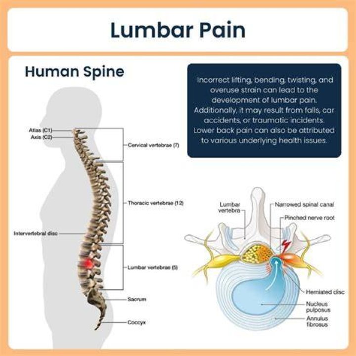
The human lower back endures exceptional daily mechanical shear forces. Five robust vertebrae, labeled L1 through L5, anchor the lumbar spine anatomy. They sit atop the sacrum, separated by intervertebral fibrocartilaginous pads that function as hydraulic shock absorbers. Each disc features a tough, fibrous exterior known as the annulus fibrosus, enclosing a gelatinous interior called the nucleus pulposus.
When you bend forward and twist simultaneously, intradiscal pressure skyrockets. If the annulus suffers micro-fissuring under that rotational shear, the gel pushes outward. This mechanical failure can manifest as a herniated disc. Adjacent to these joints run the nerve roots that converge to form the massive sciatic nerve. If structural displacement or acute chemical inflammation irritates the nerve bundle, localized back discomfort transforms into radiating sciatica nerve pain, a sharp, searing discomfort traveling down the buttock, hamstring, and calf.
Medical evaluation categorizes this neurological encroachment as radiculopathy. It introduces numbness, tingling, or diminished tendon reflexes along the affected nerve pathway. Yet, in more than 85% of clinical presentations, acute episodes stem not from structural disc rupture, but from non-specific mechanical trauma within the deep muscular stabilizers.
Inside the Violent Splint: Why Muscles Lock Tight
The most agonizing component of an acute episode rarely originates within the bone itself. It stems from the body's overprotective defense mechanism: reactive muscle strain and spasms. Deep along the posterior column run intrinsic stabilizing muscles, primarily the multifidus and the erector spinae group.
When microscopic ligament tears or facet joint capsules irritate local nerve endings, sensory neurons fire emergency distress signals directly into the dorsal horn of the spinal cord. The central nervous system perceives an acute structural threat. To prevent hypothetical spinal cord damage, the motor cortex triggers a sustained, involuntary contraction across the entire paraspinal network.
The back locks into a rigid splint. The quadratus lumborum muscles clamp shut, pulling the pelvis out of plumb and creating the telltale crooked posture seen during acute attacks. Blood flow drops within the continuously contracted tissue, causing localized hypoxia and an accumulation of metabolic waste products like lactic acid. The muscle enters a continuous ischemic spasm loop: pain causes spasm, which restricts oxygenation, triggering deeper pain.
What Scans Reveal Versus What Patients Actually Feel
The natural patient reaction to blinding lumbar pain is demanding an immediate scan. Patients assume an MRI or X-ray will pinpoint the precise defect behind their suffering. Clinical practice patterns reveal a different story. The American Academy of Family Physicians consistently advises against routine imaging within the first six weeks of non-specific back symptoms unless red-flag indicators appear.
| Imaging Modality | What It Captures Effectively | Diagnostic Limitations & Risks |
|---|---|---|
| Plain Radiograph (X-Ray) | Vertebral alignment, gross fractures, bony spurs, gross disc height collapse. | Cannot view soft tissue; blind to disc herniation, muscle strains, or nerve inflammation. |
| Magnetic Resonance Imaging (MRI) | Hydration loss, disc extrusion, neural impingement, marrow edema, soft-tissue tearing. | High false-positive rate; highlights age-related changes that may not cause the pain. |
| Computed Tomography (CT) | Complex facet joint anatomy, micro-fractures, structural bone morphology. | Exposes patient to substantial ionizing radiation; inferior soft-tissue contrast compared to MRI. |
Diagnostic imaging frequently uncovers structural anomalies that have zero correlation with the patient's immediate pain. Landmark clinical studies demonstrate that over 30% of pain-free thirty-year-olds display disc bulges on MRI scans, a figure rising to more than 60% in asymptomatic individuals over age fifty. An MRI taken during an acute flare-up often captures what radiologists term "wrinkles on the inside." Labeling these age-appropriate baseline shifts as critical defects can induce kinesiophobia, the debilitating fear of movement that paradoxically delays tissue recovery.
The Modern Treatment Playbook: From Bed Rest to Active Reloading
Decades ago, medical guidance condemned lower back pain sufferers to weeks of strict bed rest. That strategy proved disastrous. Prolonged immobilization weakens spinal musculature, stiffens connective ligaments, and prolongs neurological hypersensitivity.
Modern conservative management prioritizes rapid, active desensitization. Unless clinical red flags emerge, such as progressive lower-limb motor weakness, loss of bowel or bladder control, or suspected spinal infection, movement represents the most potent medicine available.
Prescriptive physical therapy reintroduces functional range of motion through directional preference exercises, gentle hip mobility drills, and gradual core loading. Concurrently, clinicians leverage targeted pharmacological agents, such as nonsteroidal anti-inflammatory drugs (NSAIDs) or short-term muscle relaxants, to quiet the central nervous system's threat responses. Outside the clinic, non-invasive innovations continue to expand; clinical trials supported by organizations like the Focused Ultrasound Foundation explore non-invasive sonic therapies to target stubborn nerve impingements and chronic tissue inflammation without surgical incisions.
Daily Defenses: Ergonomics, Footwear, and Dynamic Movement
Preventing an acute recurrence demands systematic adjustments to everyday physical environments. Because the spine absorbs cumulative kinetic stress from the ground upward, daily mechanical support systems play a pivotal structural role. Recent gait research highlighted by podiatric evaluations in consumer wellness outlets underscores the value of biomechanically tuned footwear. Shoes featuring moderate heel drops, structured arch contours, and firm shock-absorbing midsoles reduce ground reaction forces radiating into the lower spine during long periods of standing.
At work, static postures tax spinal ligaments far more than dynamic movement. Ergonomic support systems, including adjustable lumbar rolls, sit-stand desks, and chairs configured to preserve the natural inward lordotic curve of the lumbar vertebrae, prevent prolonged posterior disc stretching. Sleep posture demands equal diligence. Sleep Foundation research emphasizes that medium-firm mattresses deliver the optimal balance of spinal contouring and mechanical pushback, preventing the pelvis from sinking out of alignment during the night.
Frequently Asked Questions (FAQ)
Q1: What is the main difference between lumbago and sciatica?
A1: Lumbago is an overarching term referring directly to pain confined to the lower back region, typically driven by muscular, ligamentous, or facet joint irritation. Sciatica occurs when structural displacement or inflammatory chemicals irritate the sciatic nerve roots, projecting radiating, shooting pain, numbness, or weakness down the leg past the knee.
Q2: Should I go to the emergency room for an acute back spasm?
A2: Most acute back spasms do not require emergency intervention and resolve through conservative care. However, immediate emergency care is necessary if back pain coincides with red-flag symptoms: sudden loss of bladder or bowel control, numbness around the groin or saddle area, progressive leg weakness causing foot drop, high fever, or pain following major physical trauma.
Q3: How long does a standard episode of acute lower back pain take to heal?
A3: Approximately 70% to 90% of acute lower back pain episodes improve significantly within two to four weeks through active movement, over-the-counter anti-inflammatories, and physical therapy. Symptoms persisting past twelve weeks transition into chronic low back pain, requiring an expanded multimodal rehabilitation strategy.
Navigating Spinal Recovery in Modern Healthcare
An acute attack of lumbago can feel catastrophic while it unfolds. The sheer intensity of muscle contractions, paired with sharp neural irritation, often convinces individuals that their spine has suffered irreversible damage. Yet biological reality runs counter to that panic. The lumbar spine remains one of the strongest, most resilient mechanical structures in the human skeleton, built to handle massive loads and recover from acute soft-tissue trauma.
Successfully navigating an episode requires separating the intensity of the pain from the actual degree of structural tissue harm. By avoiding unneeded emergency imaging, embracing early progressive movement, addressing footwear and workstation mechanics, and working with knowledgeable physical therapists, patients regain function quickly. Understanding the mechanical cascade behind an acute back attack removes the mystery, replacing fear with an active, evidence-based plan for lasting recovery.